Our Services
Overview of Diagnostic Procedures
A complete range of neurodiagnostic and imaging procedures are available at Duke. The specific procedures needed for a patient are determined by their physician.
Imaging Studies
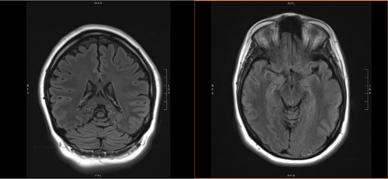
MRI
MRI can detect obvious or subtle abnormalities that may indicate a potential seizure focus. Duke offers both standard clinical MRI on 3.0 Tesla scanners as well as advanced techniques including high-resolution scans and diffusion tensor imaging.
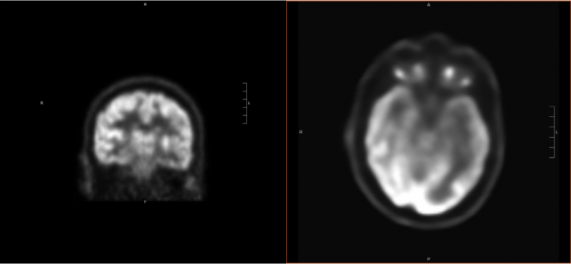
PET (Positron Emission Tomography)
PET measures the brain’s metabolic activity, which can indicate how much energy a particular part of the brain is using. Areas of the brain that are causing seizures often have different metabolic activity than the rest of the brain. The PET scan is usually performed in conjunction with an EEG to help correlate the metabolic activity with seizure activity. PET scans are typically only needed in patients who are being evaluated for epilepsy surgery.
SPECT (single-photon emission computed tomography)
SPECT may be performed during a video EEG. Like PET, this test involves the injection of a radioactive solution. It must be injected near the very beginning of a seizure, and it is only active in the body for a brief period of time. By comparing the results for an injection performed during a seizure (ictal scan) and one not during a seizure (interictal scan), it may be possible to identify the area in the brain responsible for seizures. SPECT scans typically require admission the Epilepsy Monitoring Unit.
fMRI (functional MRI)
fMRI is used to identify areas of the brain involved in certain functions. Using special MRI protocols, changes in blood flow are measured while the patient is asked to perform certain tasks. Most commonly, this is used to identify areas of the brain involved in controlling movement or language.
EEG
Routine EEG, or electroencephalography, records brain waves using electrodes placed at specific locations on the scalp using conductive paste or glue. The patient lies still on a bed and may be asked to perform some simple tasks. Flashing lights and hyperventilation may be used during the test to provide additional information. In most cases, the brain waves during both waking and sleep are recorded. The test typically takes one to two hours total time, including application and removal of electrodes.
Ambulatory EEG
An ambulatory EEG records brain waves for a longer period of time and possibly during seizures or events that are concerning for seizures. Electrodes are placed on the scalp to record brain waves, however, they are held in place with a special adhesive. The patient will go home with the electrodes and a miniature, portable recorder for one to four days. Brain waves are recorded continuously. The patient or family is asked to push a button for any events or seizures, and to maintain a detailed log of activities and events. After the device is returned to the lab, the data is analyzed.
All of our services are available to patients who book a New Patient appointment with one of our providers in the Epilepsy Clinic. For more information call 919-385-3223 or visit the DukeHealth website on epilepsy below.