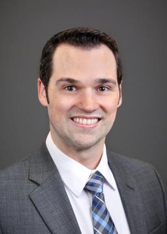
Iain Bruce, PhD, wants to get a better look at your brain. Not just at the gray, squishy lobes we’ve all seen in photographs, but at the structural changes smaller than a human hair that can lead to seizures or that eventually develop into Alzheimer’s disease. In this week’s “spotlight” interview, the new member of the Neurology Department and Brain Imaging and Analysis Center talks to us about his work advancing the field of magnetic resonance imaging (MRI) to help patients with epilepsy and other conditions. He also discusses the promise and challenges of advancing MRI technology, and how MRI may help patients receive safer, more effective surgeries in the future.
What are your responsibilities within the Neurology Department and the Brain Imaging and Analysis Center? What does a typical day for you look like?
My research responsibilities are twofold. First, I mostly work on developing new techniques for gathering, reconstructing, processing, and interpreting ultra-high spatial resolution diffusion MRI data. As the most challenging patient populations in my field are often children or adults with disorders that make it difficult to remain still for long periods of time, the overarching goal is to gather as much frequency data with the highest signal possible in the shortest amount of time. As such, a typical day often involves developing either the pulse sequences that run the scanner, the models that reconstruct the scanner signal into images, or the processing and analysis tools for interpreting the data I acquire. With that in mind, my current research project in the Epilepsy division of Neurology involves using the techniques I have developed to try and find the seizure focus in patients with intractable epilepsy - where there are no abnormalities in conventional structural MR images.
In our current studies, we are looking at cortical microstructures, and thus I am continuously working on ways to get diffusion tensor imaging data at higher spatial resolutions while preserving the signal-to-noise ratio (SNR). Once I have the data reconstructed and processed, I have been working on ways to localize abnormalities using various statistical and machine learning approaches.
How did you first get interested in brain imaging?
When I first got accepted into grad school to pursue my PhD, there happened to also be a new faculty member joining the same department as me who had a very similar background to my own. When we first met, we talked at length about his research on the statistical analysis of MR image processing operations and thus began my work and interest in brain imaging.
What do you see as the most exciting recent or pending development in your field? What questions are you hoping brain imaging is going to help us answer in the next five to 10 years?
I think the most exciting development in my field has to be the hardware improvements, such as the high-strength gradient coil we recently had installed in our scanner, that allow us to push the limits of MRI even further. With the new coil we have, the increased gradient strength has enabled us to increase spatial and temporal resolutions even further without sacrificing SNR. The results have already enabled us to characterize complex microstructures in the brain that we couldn’t see before.
While I think that these advances are a step in the right direction, I hope that the improvements we see in 10 years will be enough to push us over the edge and ultimately give way to non-invasive MRI based tools that greatly assist in pre-surgical planning. Additionally, it would be great for such advances to help produce protocols that can screen for early biomarkers associated with common diseases such as Alzheimer’s disease.
What do you enjoy most about your work?
Although most of my work involves programming, I have a background in physics and mechanical engineering, so I enjoy the process of deriving creative solutions to common problems. In a typical MRI scan, the time you have to acquire data is often short, so you have to make the most of it to get the best signal, with the highest spatial and/or temporal resolution, the highest spatial fidelity, the highest contrast, the lowest distortion, etc. It is usually a tradeoff as to which 2 or 3 of those you can realistically achieve, so I enjoy trying to come up with creative ways to get more of those features in the same amount of time.
What’s the hardest part of your job?
The most challenging part of my work is extracting meaningful information from noisy data. The higher we push the spatial resolution of images, the lower the SNR gets. SNR is proportional to voxel (3D pixels) volume, and while most MRI studies deal with data that has voxels on the order of 2x2x2 mm3, our data often has a voxel volume that can be 20 times smaller. As a result, the data we acquire often looks incredibly noisy, so it can be a challenge to derive meaningful/reliable conclusions.
What passions or hobbies do you have outside of Duke?
My wife and I have a 1-year-old son, so lately most of our hobbies involve just trying to keep up with him. Otherwise I really enjoy being outdoors, so I like activities such as hiking, fishing and traveling. We haven’t been able to do much of that over the last year, but we are looking forward to taking our son on adventures soon.

Bruce and his wife enjoy a luau in Maui during their recent honeymoon.