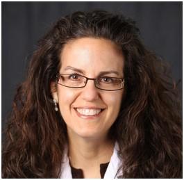
Sharon Fekrat, MD, FASRS, sees the retina, the lining of the eye that translates visual signals from the eye into what the brain perceives as sight, as both a thing of beauty and a window into the rest of the body. As a member of the Duke Departments of Ophthalmology, Neurology, and Surgery, Fekrat’s work involves treating patients with retina problems as well as leading the collaboratory iMIND (Eye Multimodal Imaging in Neurodegenerative Disease) program to understand the connections between retinal health and Alzheimer’s disease, Lewy body dementia, and other neurodegenerative disease.
For this week’s Faculty Spotlight interview, Fekrat talks to us about her multidisciplinary clinical work and research at Duke. She also shares how her recent research may allow non-invasive exams of the retina to find and help patients with early dementia with Lewy bodies years before symptoms occur. She also shares how new research and artificial intelligence will open new avenues into detecting neurodegenerative disease and other aspects of health over the next decade.
What are your current responsibilities within the Departments of Ophthalmology and Neurology? What does your typical day look like?
As a medical and surgical retina specialist who aspires to excel in all of the academic missions, every day is different. My primary appointment is in the Department of Ophthalmology, and I am fortunate to have a secondary appointment in neurology and the department of surgery. I spend my time at both Duke as Professor and the Durham VA as Associate Chief of Staff. I am involved in clinical care and take care of patients with medical and surgical retina problems in both the clinic and operating room 3 days per week. The remaining 2 days per week are devoted to the other academic missions - research, teaching/education, leadership/administration, and community.
In the clinical research space, I am Founder and Director of Duke’s iMIND – Eye Multimodal Imaging in Neurodegenerative Disease – and in this role, I lead a large research team comprised of Duke undergraduates, medical students, ophthalmology residents, and retina fellows, and I am fortunate to collaborate with numerous faculty colleagues in the Departments of Ophthalmology and Neurology as well as Computer Science and Biostatistics as well as Duke AI Health.
How and when did you first get interested in ophthalmology?
As a third-year medical student, I became fascinated with ophthalmology – the microsurgery is so precise and looking inside the eye is so beautiful. In ophthalmology, we can fix many problems with clinic-based procedures and operating room surgeries to restore sight – such an important sense and one that when intact, promotes longevity and cognitive health. When I was an ophthalmology resident, I developed an interest in the retina which lines the inside of the eyeball behind the pupil and is neurosensory tissue. The retina is the window to the rest of the body and such an important part of our bodies.
What do you enjoy most about your work?
I enjoy many aspects of my work. I enjoy helping individual patients regain some or all of their vision so that they can continue the activities in life that they enjoy. As a retina specialist, we have so many medical and surgical interventions to offer our patients.
I also enjoy the exciting iMIND research as well as mentoring and sponsoring the many students and residents and others who work with me. Above all, I enjoy making a difference and having an impact in all of the academic missions as well as thinking outside the box to move the field forward. Being a physician is an incredible privilege all around!
What’s the hardest part of your job?
The hardest part of my job is not knowing all of the answers and not being able to help every single patient. Also, the paperwork and administrative tasks continue to grow for physicians in both the clinical and research spaces - sometimes without simultaneous parallel growth of supportive infrastructure and thus physicians are not always practicing at the top of their license due to the addition of many non-physician tasks to their to-do lists. This in turn may slow down research progress and decreases access to clinical care.
You were also the lead author of a recent Journal of VitreoRetinal Diseases article examining differences in retinal and choroidal microvasculature and structure people with dementia with Lewy bodies from people with normal cognition. What were the main findings of this study and how will they help us better understand or treat dementia?
Our recent peer-reviewed publication assessing dementia with Lewy bodies (DLB) in a cross sectional study demonstrated that persons with DLB had an increased peripapillary capillary perfusion density, decreased peripapillary capillary flux index, and attenuated ganglion cell inner plexiform layer thickness compared to those with normal cognition. These findings suggest that retinal imaging metrics have the potential to serve as clinically relevant biomarkers for a diagnosis of DLB and will help us understand how these findings may differ from those with other types of dementia, such as Alzheimer's disease. We will now be imaging individuals with DLB longitudinally. We are always interested in imaging more persons with DLB!
Given that the retina is an extension of the brain, the findings we observe in the retina are also similarly occurring in the brain. It is important to be able to easily distinguish among the different causes of dementia, particularly early on in the disease process, yet we are currently unable to do so reliably with available testing. The goal of our work is to learn more about how the retina changes in these various neurodegenerative conditions over time. We hope that imaging of the retina will allow us to be able to accurately identify the various dementias before symptoms begin and earlier on in the disease process to facilitate entry into clinical trials studying novel therapeutics.
What are the biggest changes you see coming to the field of ophthalmology over the next decade?
Ophthalmology, particularly the field of retina, is a very fast moving field with rapid change. As an example, the field of retina has had at least 3 new FDA approved medications in 2023 alone. Gene therapy for common retina conditions is right around the corner and will be incorporated into clinical care in the coming years. Multimodal ophthalmic imaging is exploding and will continue to improve at an exponential pace so that the quality and resolution of structures inside the eye will continue to be unmatched.
These ophthalmic images will continue to fuel the development of AI models that will revolutionize the way we practice and may allow us to be able to diagnose systemic conditions, including neurodegenerative diseases, using retinal images and artificial intelligence. As an examination-heavy specialty, technology may eventually allow the ability to perform reliable remote examination including incorporating at-home imaging – that too is around the corner.
Ophthalmic education will incorporate virtual reality models. Our field is very fast moving with an unstoppable momentum.
What other passions or hobbies do you have outside of the Department?
I believe that we are what we eat so I have Instagram and TikTok Channels where I poke fun at processed foods with daily posts that highlight how unhealthy much of what we eat is and how most of it is sugar, fat, and salt. We are undernourished and overfed as a society.